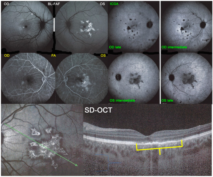
Figure 24.
APMPPE/AMIC; BL-FAF. ICGA, FA & SD-OCT findings in a case in subacute stage having received prednisone therapy for 10 days: BL-FAF (top left two frames) shows areas of hyperautofluorescence (OS > OD) corresponding to the areas of hypofluorescence (choriocapillaris non-perfusion) on ICGA (top right quartet of frames). FA (middle left two frames) shows areas of staining (OS) most probably produced by exudation of retinal vessels in response to outer retinal ischaemia. SD-OCT (bottom two pictures) shows loss and disorganisation of photoreceptor outer segments, disrupted RPE layer with overlying hyperreflective focal deposits in correspondence to hyperautofluorescence.

