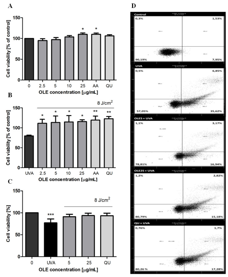
Figure 2.
The effects of different concentrations of OLE on the viability of human skin fibroblasts. Cells cultured (24 h) in the presence of OLE (2.5–25 μg/mL), vehicle (0), or the reference antioxidants AA and QU (25 μg/mL) not irradiated (A) or exposed to UVA-irradiation (8 J/cm2) (B) were cultured for an additional 24 h, and then subjected to CCK-8 assay. The cytotoxicity of OLE and fibroblast survival were expressed as a percentage of control, where the absorbance of untreated and of corresponding non-irradiated cells were taken as 100%, respectively. The post-radiation viability of fibroblasts evaluated by the Annexin V-FITC/PI staining (flow cytometry) assay (C). A representative dot-plot histogram is shown (D), cell populations in II, I and III/IV quadrants represent alive, undergoing necrosis and undergoing apoptosis Hs68 cells, respectively. The figure shows mean results from 3–6 independent experiments. Error bars denote ±SD. * p < 0.05; ** p < 0.01; *** p < 0.001 compared to control (0).

