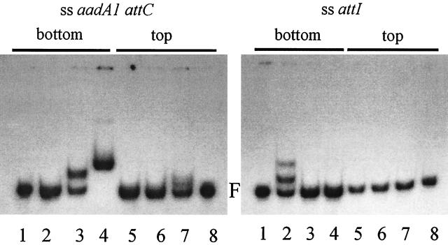
FIG. 4.
Gel retardation assays with ssDNA fragments containing either the top or the bottom strands of the aadA1 attC site (left) or the attI site (right). The substrate DNA used is indicated at the top of the figure. Protein extracts in each lane are as follows: left panel, lane 1, control DNA; lane 2, E. coli C41 control extract; lane 3, IntI1-COOH; lane 4, IntI1; lane 5 E. coli C41 control extract; lane 6, IntI1-COOH; lane 7, IntI1; lane 8, control DNA; right panel, lane 1, DNA alone; lane 2, purified IntI1; lane 3, IntI1-COOH; lane 4, E. coli C41 control extract; lane 5, IntI1; lane 6, IntI1-COOH; lane 7, E. coli C41 control extract; lane 8, control DNA. F, free DNA.

