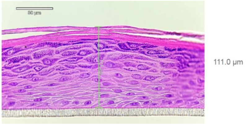
Figure 2.
Histology report on EpiDerm tissue (HRE, EPI-200; LotNo:36125)) after hematoxilin-eosin staining provided by MatTek Life Sciences. Well-differentiated epidermis consisting of basal, spinous, granular layer of keratinocytes and Stratum corneum can be seen. At least 4 viable cell layers are present. The tissue thickness is 70–130 µm (average 111.0 µm).

