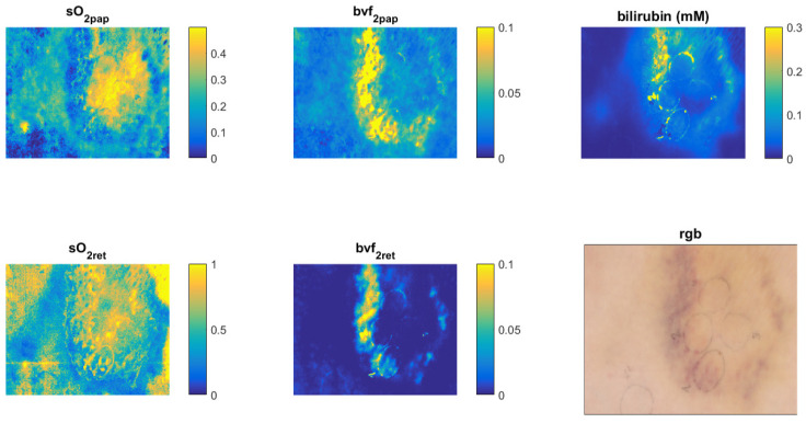
Figure 14.
An example of utility of the custom-made laboratory hyperspectral imaging (HSI) system, with shown tissue property maps of a skin bruise extracted from a hyperspectral image using the Inverse Diffuse Approximation Algorithm. The presented tissue parameters are: sO2pap—blood oxygenation in the papillary dermis, sO2ret—blood oxygenation in the reticular dermis, bvf2pap—blood volume fraction in the papillary dermis, bvf2ret—blood volume fraction in the reticular dermis, bilirubin (mM)–bilirubin concentration in the dermis, and RGB—an RGB image of the bruised skin. All quantities are fractions without units, except bilirubin, which has millimolar units (mM).

