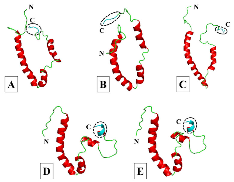
Figure 3.
Cartoon representation of the three-dimensional (3D) models of the SARS-CoV-1, -2, MERS-CoV, HCoV-229E, and HCoV-NL63 envelope (E) proteins. (A) SARS-CoV-1. (B) SARS-CoV-2. (C) MERS-CoV. (D) HCoV-229E. (E) HCoV-NL63. Models were generated using MODELLER software and based on the partially resolved nuclear magnetic resonance (NMR) structures for SARS-CoV-1 E (PDB IDs: 5X29 and 2MM4) obtained from the Research Collaboratory for Structural Bioinformatics (RCSB) protein data bank (PDB) [49,50,51,52]. The models for SARS-CoV-1, -2 and HCoV-229E were generated from the 5X29 template, the MERS-CoV model was constructed from the 2MM4 template, and the HCoV-NL63 model was generated using the HCoV-229E E protein model as the template. The amino (N) and carboxy (C)-termini are indicated accordingly; coils are shown in green, α-helices in red, and the C-terminal PDZ-binding motif (PBM) in cyan and enclosed by a circle of dashed lines.

