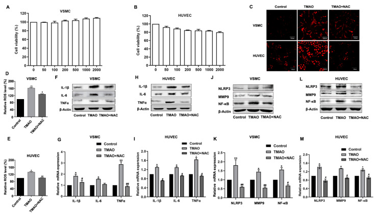
Figure 1.
TMAO-induced inflammation via ROS in VSMCs and HUVECs. (A,B). VSMCs (A) and HUVECs (B) were incubated with different concentrations of TMAO (100, 200, 500, 1000 and 2000 μM) for 24 h. Thereafter, the cell viability was determined. (C–E) VSMCs and HUVECs were treated with 1000 μM TMAO for 24 h. The total ROS levels were determined by staining with DCFH-DA. Then, the ROS levels were observed by confocal microscopy (C) and measured by flow cytometry (D,E). (F–M) The protein levels (F,H,J,L) and relative mRNA levels (G,I,K,M) of inflammation-related markers IL-1, IL-6 and TNFα (F–I) and other inflammatory factors NLRP3, MMP9 and NF-κb (J–M) in VSMCs (F,G,J,K) and HUVECs (H,I,L,M) were detected by western blot and quantitative real-time PCR. The values are the mean ± SD; n = 3; one-way ANOVA followed by a Tukey post hoc test. * p < 0.05; ** p < 0.01 vs. Control; # p < 0.05; ## p < 0.01 vs. TMAO group.

