
Figure 1.
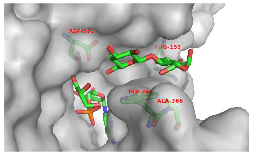
The amino acid residues in the lactose-binding pocket of Psp2,6ST(15-501)-His6. The protein structure was modeled based on the reported crystal structure of Δ16pspST6 (pdb: 2Z4T) and analyzed using PyMOL (carbon, nitrogen, oxygen, and phosphorus atoms in lactose and the amino acid residues of interests (stick model) are shown in green, blue, red, and orange colors, respectively).