
Fig 6. Mice with a “humanized” non-IBD virome had attenuated intestinal inflammation while those with a “humanized” IBD-derived virome exhibited intestinal inflammation.
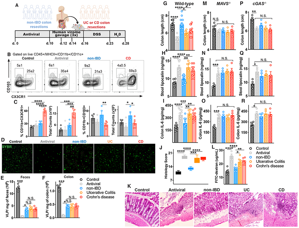
(A) Schematic of mouse gut virome depletion using an antiviral cocktail (acyclovir 20 mg/kg, lamivudine 10 mg/kg, ribavirin 30 mg/kg, oseltamivir 10 mg/kg, gray, daily gavage for 10 days), and reconstitution with 200 μL of human non-IBD or IBD VLPs pool (4×10⁸ VLPs per mouse) by gavage on day 10, 12 and 14. 2.5% DSS colitis model was commenced on day 16. (B) Flow cytometry plots of CD45+MHCII+CD11c+CD11b+ cells from the colonic lamina propria, and percentages and counts of (C) CX3CR1+ or CD103+ mononuclear phagocytes in control, antiviral depleted (AV), non-IBD or IBD humanized virome mice on day 17. (D-F) Confocal images and quantification of viral-like particles (VLPs) using SYBR Gold in feces or colon tissue of humanized virome mice. (G) Colon length, (H) stool lipocalin, (I) IL-6 levels in colonic explant supernatants cultured for 24 hours following 12 days of induced DSS colitis measured by ELISA. (J,K) Representative hematoxylin and eosin–stained sections and blinded histologic scores in mouse colon tissue (scale bar, 50 μm). (L) FITC-dextran level in serum 4 hours after oral gavage following 7 days of DSS. (M-O) Colon length, stool lipocalin or colonic explant IL-6 levels following induction of DSS colitis in antiviral-treated MAVS−/− mice or (P-R) cGAS−/− mice administered human non-IBD, UC or CD colon-derived viromes. Data are mean ± s.e.m. of 4-8 animals. Representative of 2 independent experiments. *P<0.05, **P<0.01, ***P<0.001, ****P<0.0001, two-tailed unpaired t-test or one-way ANOVA with Tukey’s multiple comparison test.