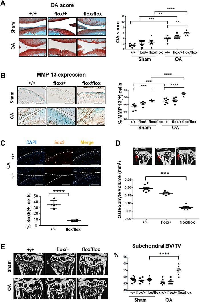
Fig. 1. Lin28a-deficient chondrocytes exacerbate cartilage degradation in mice with OA.
Ablation of Lin28a in chondrocytes was induced in Lin28atm1.2Gqda/Col2a1-CreER knockout homozygous (flox/flox) and heterozygous (flox/+) mice by intraperitoneal tamoxifen injection in 9-week-old mice; Col2a1-CreER (+/+) littermates were controls (CTs). OA was induced at age 10 weeks, and then mice were euthanized 8 weeks after OA induction and analyzed at age 18 weeks. (A) Safranin-O staining of sham and OA joints (scale bars, 100 μm). Graph represents OA score in sham and OA joints. (B) Immunohistochemistry of MMP13 content (scale bars, 100 μm). Graph represents the percentage of MMP13-positive cells in sham and OA mice. (C) Immunofluorescence of SOX9 in OA mice (scale bars, 200 μm). Graph represents the percentage of Sox9-positive cells. (D) Osteophyte volume analyzed by microtomography in OA mice. (E) Subchondral bone volume to total volume (BV/TV) analyzed by microtomography in sham and OA mice and quantification. Data are means ± SEM. **P < 0.01, ***P < 0.005, and ****P < 0.001.

