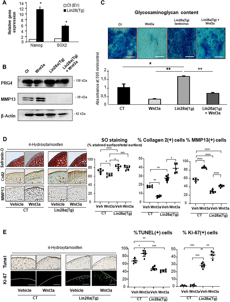
Fig. 2. Lin28a overexpression increased extracellular matrix production in vitro.
Wild-type mouse primary chondrocytes were transduced with Lin28a lentivirus [Lin28a(TG)] or empty vector [Ct (EV)] and cultured for 48 hours or 1 week at 1% O2 in the presence of Wnt3a conditioned medium (Wnt3a-CM) to trigger chondrocyte catabolism. (A) RT-qPCR analysis of mRNA levels of stemness genes. (B) Western blot analysis of PRG4 and MMP13 protein expression in primary chondrocytes transduced with Ct (EV), Ct (EV), with Wnt3a-CM (wnt3a), Lin28a(TG), and Lin28a lentivirus + Wnt3a-CM [Lin28a(TG) + Wnt]. (C) Alcian Blue staining and spectrophotometry quantification of sulfated glycosaminoglycans in primary chondrocytes after 1 week of culture (scale bars, 200 μm). Femoral explants were harvested from 10-week-old TgLin28aflox/flox/Col2a1-CreER [Lin28a(Tg)] or Col2a1-CreER (CT) mice and treated with Wnt3a-CM (Wnt3a) or not (vehicle). 4-Hydroxitamoxifen was used to induce in vitro recombination. (D) Safranin-O staining and immunohistochemistry (collagen 2, MMP13) of CT and Lin28a(Tg) explants (scale bars, 100 μm). Graphs show the percentage of ratio of Safranin-O–unstained cartilage to Safranin-O–positive cartilage and the percentage of MMP13-positive and collagen 2–positive cells. (E) Apoptosis was assessed by terminal deoxynucleotidyl transferase–mediated deoxyuridine triphosphate nick end labeling (TUNEL) assay and proliferation by Ki-67 immunofluorescence staining. Data are means ± SEM. *P < 0.05, **P < 0.01, ***P < 0.005, and ****P < 0.001.

