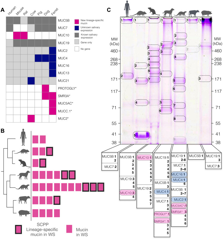
Fig. 5. Comparison of mucins in saliva of diverse mammalian species.
(A) Mucin proteins in whole saliva of different mammalian species identified by LC-MS analysis. Mucins previously not known to be expressed in saliva are colored in dark blue boxes. Lineage-specific mucins identified in this study are boxes in magenta. Gray boxes indicate mucins with previously known salivary expression, while light gray indicates that the gene is present in the species’ genome with no expression detected in saliva. Empty boxes indicate that the species does not have the corresponding gene. Gene annotations were used as provided by the respective assemblies. Longer gene names indicated by an asterisk were shortened (PROGLY: PROTEOGLYCAN-LIKE; MUC2: MUC2-LIKE; MUC5AC: MUC5AC-LIKE; MUCC.1: MUCC.1-LIKE). (B) Graphical representation of the data in (A) to indicate the total number of mucin proteins expressed in whole saliva (WS) of human, mouse, rat, dog, ferret, cow, and pig (magenta rectangles). Lineage-specific mucins found within the SCPP locus are indicated by a black border. (C) Whole saliva of the above mammalian species separated by SDS-PAGE and stained with periodic acid–Schiff to reveal glycosylated proteins. Gel bands that were analyzed by LC-MS are circled. Gray circles indicate bands where a mucin could not be identified. Vertical banners below the gel lanes show the identified mucins with numbers corresponding to bands in the gel. Magenta highlights indicate lineage-specific orphan mucins, while blue highlights indicate known mucin proteins that were not previously identified in saliva. MW, molecular weight.

