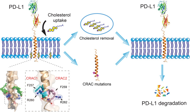
Fig. 6. Schematic diagram depicting the effect of cholesterol on PD-L1 stability.
Cholesterol can directly bind to the transmembrane domain of PD-L1 at two CRAC motifs, stabilizing PD-L1 on the cell membrane. Reducing cholesterol levels with a 3-hydroxy-3-methylglutaryl coenzyme A (HMG-CoA) reductase inhibitor (simvastatin) or cholesterol depletion reagent (MCD) decreases levels of membrane-bound PD-L1 by promoting PD-L1 ubiquitination and degradation. Mutations in the CRAC motifs have similar effects, reducing levels of PD-L1 on the cell surface. The magnified region shows key residues (CRAC1: F257/R260; CRAC2: F259/R262) in the two cholesterol-binding sites of PD-L1-TC.

