Figure 1.
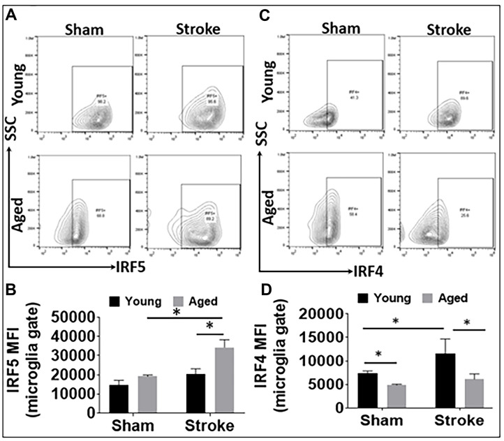
IRF5 and IRF4 expression levels in microglia in young and aged C57BL/6 mice. (A, B) Representative flow plots of microglial IRF5 (A) and the mean fluorescence intensity (MFI) (B). (C, D) Representative flow plots of microglial IRF4 (C) and the MFI) (D). n = 4-5 /group sham and 6-7/group stroke; *p<0.0500.

