
Extended Data Figure 7. STING-induced LC3 conversion does not require Beclin-1 (BECN1) or VPS34.
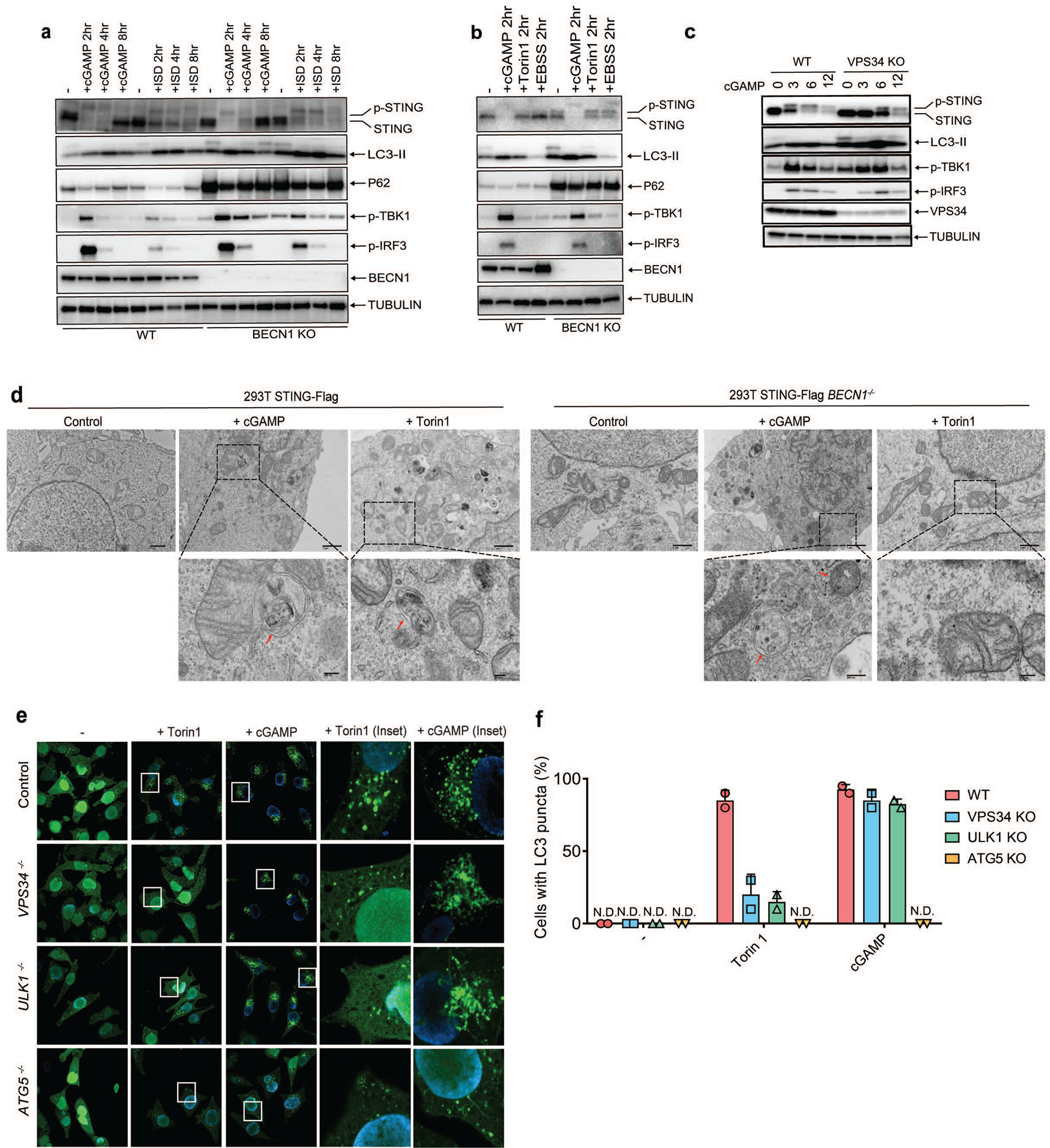
a, BECN1 is dispensable for LC3 conversion triggered by cGAMP. Wild type and BECN1−/− BMDM were stimulated with cGAMP or HT-DNA at the indicated time followed by immunoblotting of cell lysates. b, BECN1 is not essential for LC3 conversion in conventional autophagy. Wild type and BECN1−/− BMDM were stimulated with cGAMP or Torin 1 or cultured in EBSS starvation media at the indicated time followed by immunoblotting of cell lysates. c, VPS34 depletion delayed cGAMP-induced STING degradation but not LC3 lipidation. VPS34 knock-out BJ cells were treated with cGAMP for the indicated time followed by immunoblotting of cell lysates. d, Electron micrographs of 293T STING-Flag and 293T STING-Flag BECN1−/− cells, stimulated with cGAMP or Torin1. Boxed areas are enlarged to show double-membrane organelles that represent autophagosomes. Red arrow highlights double-membrane characteristic of autophagosomes in stimulated cells. Scale bar is 1 μm for original picture and 200 nm for the zoomed pictures. e&f, ULK1 and VPS34 are essential for LC3 puncta formation induced by Torin 1 but not by cGAMP. ULK1−/−, VPS34−/−, or ATG5−/− Hela GFP-LC3 cells were treated with Torin 1 or cGAMP for the indicated time. GFP-LC3 puncta formation was visualized by fluorescence microscopy (e) and the percentage of cells with GFP-LC3 peri-nuclear foci formation was quantified (f). N.D., not detectable. The percentage of cells with LC3-GFP puncta was quantified from 100 cells (n = 2).