. 2022 Aug 15;11:e80148. doi: 10.7554/eLife.80148

Figure 4. E17K and Y18A mutations promote the catalytic activity of Akt and disrupt the interdomain interactions between the PH and kinase domains.

(A) MST (microscale thermophoresis) binding experiments using N-terminally Cy5-labeled kinase domain with pT308 as a target protein and isolated PH domain as a ligand. R86A: blue, E17K/R86A: yellow, Y18A/R86A: red (n=3, SEM shown). (B) Steady-state kinetic plots of full-length Akt mutants with pT308 (B) in the absence or (C) presence of a pS473 C-tail modification (A6–A13). Kinase assays were performed with each Akt mutant in buffer containing 20 μM GSK3 peptide substrate and varying amounts of ATP (0–2.5 mM). Kinetic parameters of each Akt mutant were determined from V/[E] versus [ATP] plots (n=2, SEM shown).

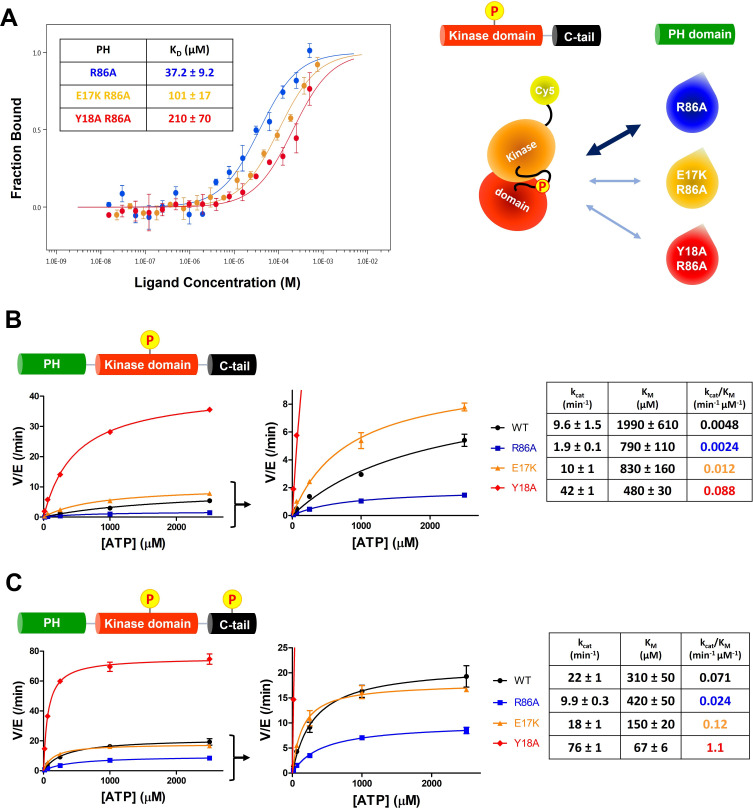
Figure 4—source data 1. Kinase activity assays with full-length Akt mutants (WT, R86A, E17K, Y18A) having pT308 and non-phosphorylated C-tail.
Figure 4—source data 2. Kinase activity assays with full-length Akt mutants (WT, R86A, E17K, Y18A) having pT308 and pS473.

Figure 4.

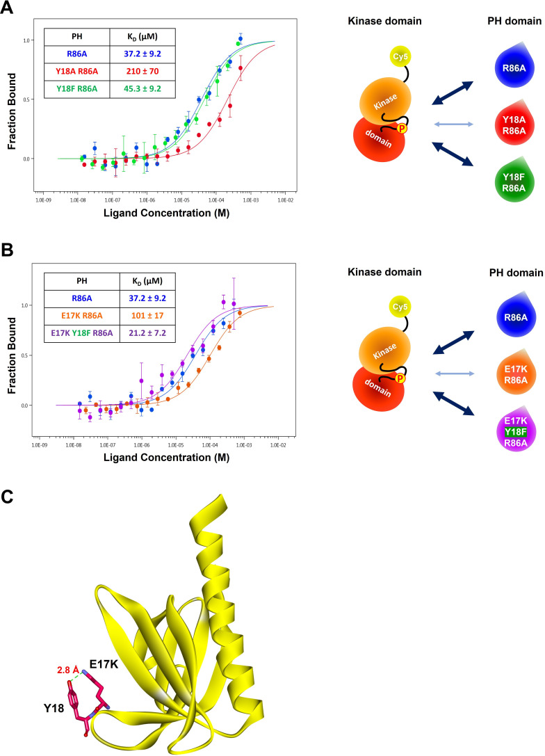
Figure 4—figure supplement 1. The role of the hydroxyl group of Tyr18 in the PH-kinase domain interaction.

Figure 4—figure supplement 1.

(A–B) MST (microscale thermophoresis) binding experiments using N-terminally Cy5-labeled kinase domain with pT308 as a target protein and isolated PH domain as a ligand. R86A: blue, Y18A R86A: red, Y18F/R86A: green, E17K/R86A: yellow, E17K/Y18F/R86A: purple (n=3, SEM shown). The obtained binding affinity (KD) values were showed in the tables. (C) Crystal structure of the E17K PH domain complexed with IP4 (PDB: 2UZS) showing a hydrogen bond between the mutated lysine residue at the Glu17 position and the hydroxyl group of Tyr18 (IP4, not shown).
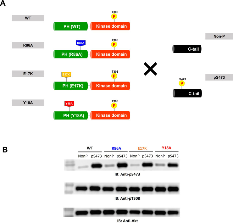
Figure 4—figure supplement 2. Semisynthesis of Akt mutants containing pT308 in the absence or presence of pS473 using a two-piece ligation strategy.

Figure 4—figure supplement 2.

(A) Semisynthesis of Akt mutant forms having pT308 with or without pS473 (A6–A13). The intact PH domain-kinase domain constructs having different mutations were expressed using insect cells and then ligated with either non-phosphorylated or S473-phosphorylated synthetic C-tail peptides to make full-length Akt mutant forms. PDK1-catalyzed phosphorylation was performed to introduce pT308 into the activation loop before the C-tail ligation reaction. (B) Western blot analysis to confirm the phosphorylation levels at Ser473 and Thr308 of each Akt construct.
Figure 4—figure supplement 2—source data 1. Raw western blot images showing the phosphorylation levels at Ser473 and Thr308 of each Akt construct.
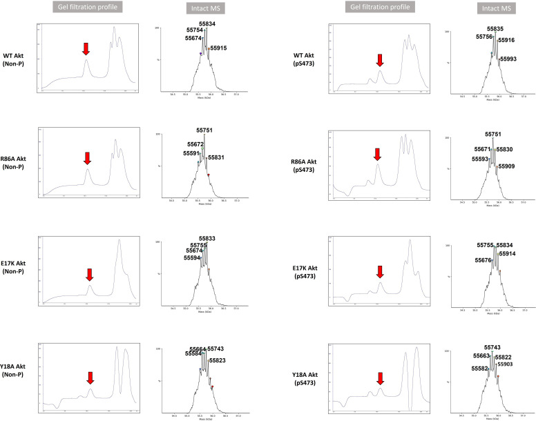
Figure 4—figure supplement 3. Validation of full-length Akt mutant proteins.

Figure 4—figure supplement 3.

Gel filtration profile and intact MS analysis of each full-length Akt mutant having activation loop phosphorylation at Thr308 with or without C-tail phosphorylation at Ser473 (Non-P or pS473). Calculated molecular weight of each Akt construct assuming Thr450 is phosphorylated and its N-terminus is acetylated after the N-terminal methionine cleavage: WT(Non-P) (A6): 55758.5 Da, R86A(Non-P)(A7): 55673.4 Da, E17K (Non-P)(A8): 55757.6 Da, Y18A(Non-P)(A9): 55666.5 Da, WT(pS473)(A10): 55838.5 Da, R86A(pS473)(A11): 55753.4 Da, E17K (pS473)(A12): 55837.6 Da, Y18A(pS473)(A13): 55746.4 Da.