. 2022 Aug 15;11:e80148. doi: 10.7554/eLife.80148

Figure 5. Phospholipid, phosphatidylinositol 3,4,5-triphosphate (PIP3) binding affinities and PP2A-mediated phosphatase removal of pT308 among several full-length Akt mutant forms.

(A) PIP3 binding assays with several isolated Akt PH domain forms. WT: black, R86A: blue, E17K: yellow, Y18A: red, E17K Y18A: green. Fluorescent anisotropy spectra were obtained from the mixture of 50 nM fluorescein-labeled soluble (C8) PIP3 and varying amount of Akt PH domain (0–50 μM). The KD values were determined from fluorescence anisotropy versus PH domain protein concentration plots (n=3, SD shown). (B) Full-length Akt mutants have different sensitivities toward the dephosphorylation of pT308 by PP2A phosphatase. Dephosphorylation assays were performed with Akt mutants containing pT308 and lacking C-tail phosphorylation (A6–A9). The dephosphorylation rates were monitored by western blots with anti-pT308 antibody and T1/2 values shown were determined from plots for fraction of pT308 versus time (n≥3, SEM shown).

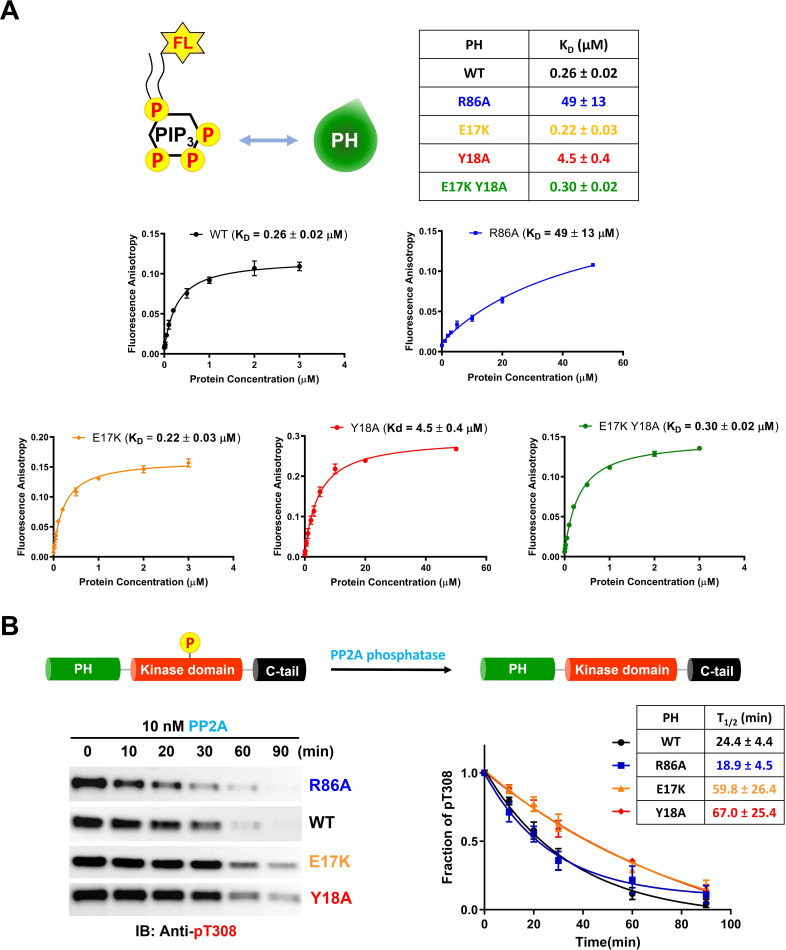
Figure 5—source data 1. Fluorescence anisotropy binding experiments to measure the phospholipid, phosphatidylinositol 3,4,5-triphosphate (PIP3) binding affinities of the PH domain mutants.
Figure 5—source data 2. Dephosphorylation assays using PP2A phosphatase on full-length Akt mutants containing pT308 and a non-phosphorylated C-tail with raw western blot images.

Figure 5.

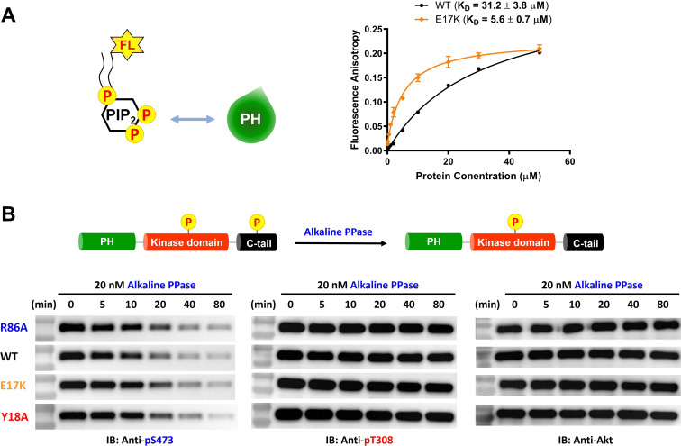
Figure 5—figure supplement 1. E17K mutation on the PH domain enhances its phosphatidylinositol 4,5-bisphosphate (PIP2) binding affinity while not changing sensitivity toward dephosphorylation of pS473.

Figure 5—figure supplement 1.

(A) PIP2 binding assay with the isolated PH domain. WT: black, E17K: yellow. Fluorescent anisotropy spectra were obtained from mixtures of 50 nM fluorescein-labeled soluble (C8) PIP2 and varying amounts of Akt PH domain (0–50 μM). The KD values were determined from plots of fluorescence anisotropy versus PH domain protein concentration (n≥3, SD shown). (B) Alkaline phosphatase dephosphorylates Akt mutants at pS473 with similar rates, while not dephosphorylating pT308 under these conditions. Akt mutants with pT308 and pS473 (A10–A13) were employed for dephosphorylation assays with alkaline phosphatase. The dephosphorylation rates were monitored by western blots with anti-pS473, anti-pT308, anti-Akt antibodies.
Figure 5—figure supplement 1—source data 1. Fluorescence anisotropy binding experiments to measure the phosphatidylinositol 4,5-bisphosphate (PIP2) binding affinities of the PH domain mutants.
Figure 5—figure supplement 1—source data 2. Dephosphorylation assays using alkaline phosphatase on full-length Akt mutants having pT308 and pS473 with raw western blot images.