. 2019 Sep 26;1(10):4137–4147. doi: 10.1039/c9na00328b

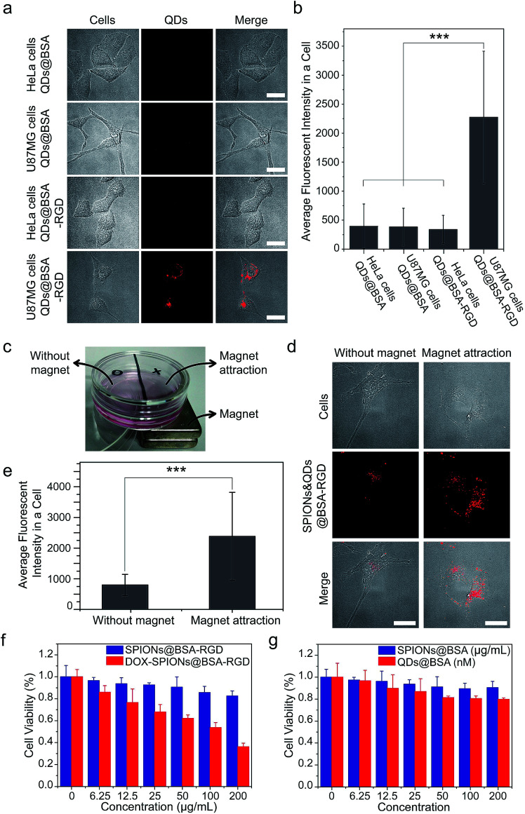
Fig. 6. Proof-of-concept studies of biological applications of supraparticle co-assemblies. (a and b) QDs@BSA were conjugated with the RGD peptide, and the formed QDs@BSA-RGD were able to specifically label and image U87MG cells. (a) Shows representative images. (b) Shows quantification results using the images obtained from the experiments of (a). Images of ∼100 cells from each sample were used for the quantification. ***P < 0.001. (c–e) The bi-functionality of SPIONs&QDs@BSA-RGD could be used to achieve combined magnetic targeting and fluorescence imaging of biological cells. (c) Shows the experimental setup. U87MG cells were cultured on the bottom of the dish. SPIONs&QDsBSA-RGD dispersed in the cell culture medium were incubated with the cells. (d) Shows representative images. (e) Shows quantification results using the images obtained from the experiments of (d). Images of ∼100 cells from each sample were used for the quantification. ***P < 0.001. (f) An anticancer drug doxorubicin (DOX) was loaded onto supraparticle co-assemblies to achieve cancer cell-killing effects. The DOX-loaded supraparticle co-assemblies were freshly prepared for the cancer cell-killing experiments (storage time < 1 day). (g) Cell viability studies show that supraparticle co-assemblies are biocompatible.

Fig. 6