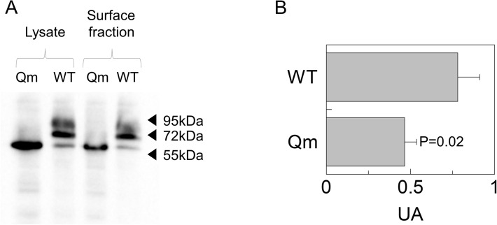
Figure 4.
Cell surface biotinylation of CD98. Surface protein fractions isolated as described in material and methods were entirely loaded on SDS-PAGE for western blot analysis; total lysate used to perform membrane protein fraction isolation was shown as a control. Loading order: Qm total lysate (lane1), WT total lysate (lane 2), Qm surface fraction (lane 3) and WT surface fraction (line 4). The western blot was performed using anti-FLAG33. The image is representative of three independent experiments. The histogram represents mean of values obtained by scanning densitometry of immunoblots. Statistical analysis was performed for each construct measuring membrane fraction with respect to the total lysate by Student's t-test.

