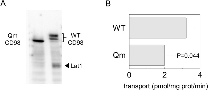
Figure 7.
Cell surface biotinylation of CD98 and LAT1. In (A), Surface protein fractions isolated from HEK293 transfected with WT-CD98 (lane1) or Qm-CD98 (lane 2) constructs as described in material and methods. The obtained samples were tested for the presence of native LAT1 using an anti-LAT1 antibody18. WT and Qm mutant were also tested to ensure that sufficient amount of membrane protein fraction was loaded in the gel using anti-FLAG antibody33. The western blot image is representative of three independent experiments. In (B), Uptake of [3H]-histidine in HEK293 cells. HEK293 cells were seeded and transfected with WT-CD98 or Qm-CD98 constructs as described in materials and methods; transport was started by adding 5 μM [3H]-histidine to transfected cells. The transport was blocked after 1 min, by placing cells on ice and washing them with cold transport buffer as described in materials and methods. Results are means ± SD of three independent experiments. Significantly different from WT as estimated by Student's t-test.

