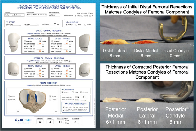
Fig. 1.
Composite of images shows the intraoperative verification worksheet of a patient including entries for patient number, surgeon name, sex, age, BMI, time to complete corrected femoral cuts, date of surgery, right or left knee, type of primary deformity (varus, valgus, or patellofemoral), condition of ACL, plan thickness of distal medial and lateral and posterior medial and lateral femoral resections, initial and corrected caliper thickness of each femoral resection (left) and the recordings of the thicknesses of the distal and posterior femoral bone resections compared to the thicknesses of the condyles of the femoral component (right)

