FIG. 2.
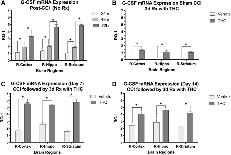
Effects of THC treatment on G-CSF mRNA expression in brain regions on the side of injury (right hemisphere; n=4 mice per group). X-axis=brain regions, Y-axis is change in mRNA expression (RQ-1). (A) G-CSF expression increased in the three brain regions measured in a time-dependent manner after CCI in untreated mice. Two-way ANOVA revealed that time contributed more than brain region to total variance; Sidaks correction for multiple comparison showed significant differences between times after CCI in each region (*<0.001); (B) THC treatment for 3 days in animals that had sham surgery (no CCI) also resulted in increased mRNA expression of G-CSF in the three brain regions measured on day 3. Two-way ANOVA revealed that time, but not brain region contributed significantly to total variance. G-CSF mRNA in each brain region was significantly increased by THC compared to vehicle treatment (*p<0.001); (C, D) G-CSF mRNA was also significantly increased by THC treatment compared to controls in the three brain regions on days 7 and 14 after CCI. Two-way ANOVA revealed that treatment contributed more than brain region to total variance. Sidak's correction for multiple comparisons showed that all three brain regions had significantly increased G-CSF mRNA upregulation on days 7 and 14 (*p<0.05). G-CSF, granulocyte-colony stimulating factor; RQ-1, relative quantification-1.

