. 2019 Aug 21;1(10):3980–3991. doi: 10.1039/c9na00289h

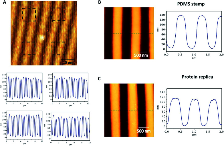
Fig. 4. AFM characterization of the nanostructured protein films. For a PDMS stamp (A) and the protein replica (B), AFM images of 2 × 2 μm2 and a height profile are shown. The cross-section of the replica shows a perfect replication of the nanopatterns on the protein film, with a height of approximately 140 nm and a periodicity of 700 nm (C). The top panel shows a 50 × 50 μm2 image acquired by AFM, in which the film patterns can be observed. The black squares show the four areas of 10 × 10 μm2 that were selected to evaluate the topography of the surface. The bottom panels show the AFM profiles of the four areas selected, revealing a regular topography, with heights around 140 nm.

Fig. 4