. 2022 Jun 7;4(14):3054–3064. doi: 10.1039/d2na00169a

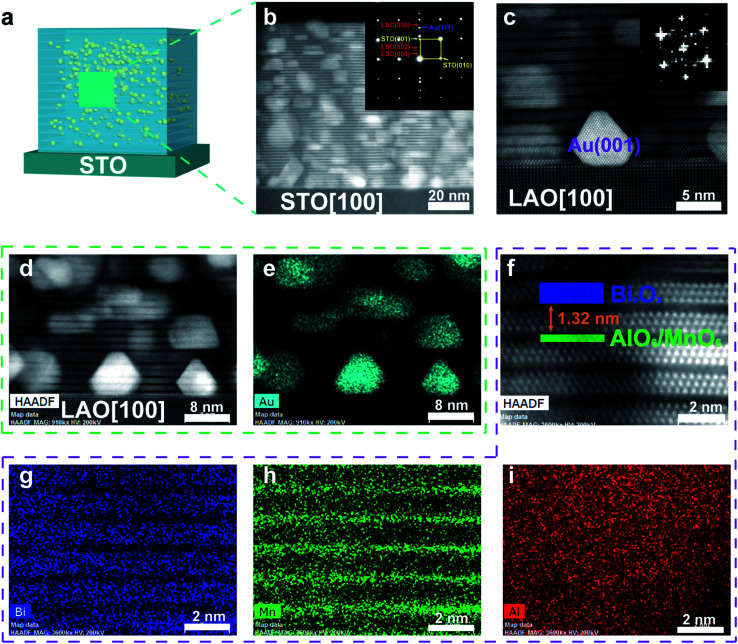
Fig. 2. Microstructure characterization of BAMO-Au thin films. (a) A schematic drawing of the BAMO-Au thin film. (b) High-resolution STEM image of the BAMO-Au thin film taken along the STO[100] zone axis. The inset shows the SAED pattern of the thin film and the substrate. (c) High-resolution STEM image of the BAMO-Au thin film taken along the LAO[100] zone axis. Au NPs are (001)-oriented at the interface between the substrate and the film. (d) High-resolution STEM HAADF image and (e) EDS map of the Au NPs embedded in the matrix. (f) High-resolution STEM HAADF image and (g, h and i) EDS maps together show the layered structure of the BAMO matrix.

Fig. 2