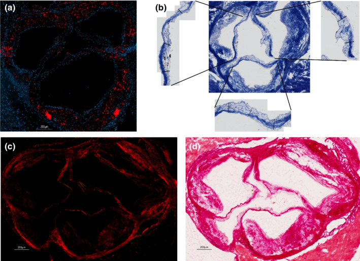
FIGURE 2.

Aortic valve histology methods. (a) Shows the DAPI (4',6‐diamidino‐2‐phenylindole ‐ bleu) coloration of cell nucleus and the OsteoSens* fluorescence (red) of the calcium deposits. (b) Shows the Trichrome Masson staining and the measurement of each leaflet. (c and d) Show the Picrosirius red staining under polarized (c) and white (d) light.
