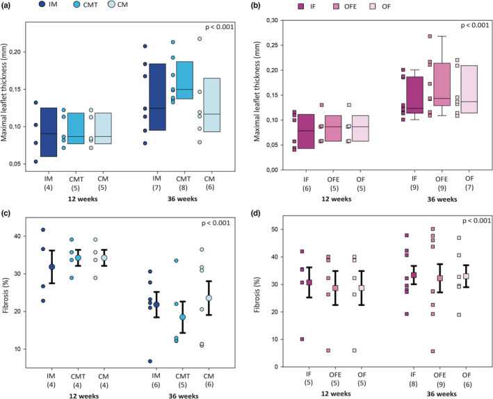
FIGURE 4.

Aortic valve staining presenting valve thickness and fibrosis for each group, at 12 and 36 weeks of age. Average maximal thickness of each aortic valve leaflet was evaluated after Masson Trichrome staining in each group of male (a) and female (b) mice. Percentage of fibrosis was evaluated by red picrosirius staining in each group of male (c) and female (d) mice. Legend as in Figure 1.
