. 2021 Oct 25;3(24):6762–6796. doi: 10.1039/d1na00447f

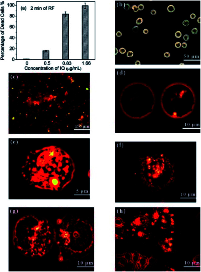
Fig. 14. (a) Percentage of dead cells as the function of IQ concentrations under the RF treatment of 2 min. Confocal microscopic images of (b) normal Panc-1 cells, (c) IQ nanocomposite incubated with Panc-1 cells for 24 h. (d) Nanocomposites (without and with UV radiation) were uptaken into the cell cytoplasm, and they were found to agglomerate around the nuclear membrane, and a small number of them crossed the nuclear membrane into the nucleus. (e–h) Cell shrinkage, membrane blebbing, the disintegration of the nuclear membranes, and apoptotic bodies as they resulted from the RF heating process. Reproduced from ref. 5 with permission from American Chemical Society, copyright 2010.

Fig. 14