. 2021 Oct 25;3(24):6762–6796. doi: 10.1039/d1na00447f

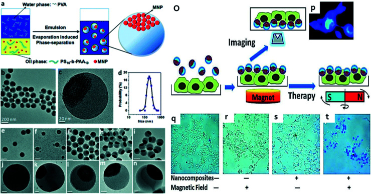
Fig. 18. (a) Schematic illustration of the preparation of multifunctional nanocomposites with spatially separated functionalities. (b and c) TEM images of the nanocomposites at different magnifications (d) hydrodynamic size (e–n) with increasing concentrations of MNPs, the size of the nanocomposites increased and their magnetic segments. Scale bars: (e–i) 200 nm and (j–n) 20 nm. (o) Schematic illustration of the experimental conditions and magnetolytic therapy. Nanocomposites are first brought down to cell surface by magnetic attraction. Cells can then be examined by fluorescence microscopy or treated with oscillating magnetic fields, which result in mechanical forces to break down cell membranes. (p) Fluorescence imaging of cells coated with nanocomposites. (q–t) Magnetolytic therapy on tumor cells. Reproduced from ref. 247 with permission from American Chemical Society, copyright 2010.

Fig. 18