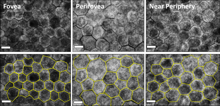
Figure 2.
RPE cell monolayer at three locations (fovea, perifovea, near periphery) of healthy donors with and without marked cell borders. RPE flatmounts imaged with a confocal fluorescence microscope (excitation: 488 nm) at three predefined locations. The tissues are from healthy donors: 81 years, 85 years, and 83 years old, respectively (from left to right). Bottom row: the RPE cell borders are delineated in yellow. The image clearly demonstrates the RPE cell size differences between the fovea and extrafoveal locations. Also, due to high lipofuscin granule load, perifoveal RPE cells appear to have the highest autofluorescence signal compared to foveal and near-peripheral RPE cells. Scale bar: 10 µm.

