. 2021 Feb 19;3(9):2466–2474. doi: 10.1039/d0na01027h

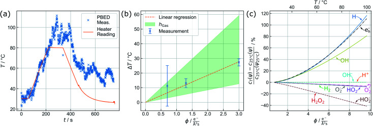
Fig. 3. (a) Active heating protocol of a TEM liquid cell (orange) and in situ temperature profile acquired from parallel beam electron diffraction by Au nanoparticles (x) in the observation area. Particle-based measurements perfectly reproduce the temperature ramp (until 220 s) but show an offset related to beam heating effects upon subsequent delay and decline. (b) PBED-derived increase of specimen temperature after 5 min of illumination depending on the electron flux density ϕ. Error bars mark the maximum deviation of the {220} and {311} data from the mean value. The green area spans the considerable range of temperature variation modelled for a gaseous environment (see text). (c) Beam heating strongly affects the equilibrium concentrations of water radiolysis products and thus the prospected chemical environment. Assuming a reference temperature of 25 °C, the curves give the percental change of relevant species at the corresponding dose rate ψ against a conventional radiolysis model neglecting beam-heating effects. The corresponding sample temperature in the observation area is plotted on the top axis.

Fig. 3