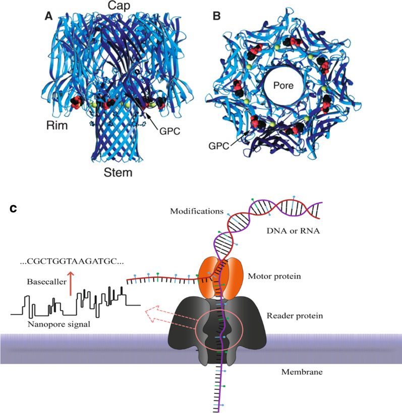
Fig. 4.
Representation of α-hemolysin from Staph aureus. Reprinted from [91] with permission of the publisher (CCC License ID: 5196920847168, 27 Nov 2021). A Side view of the alpha-hemolysin heptameric complex indicates the exact location of the phospholipid bilayer. B View of alpha-hemolysin from the cis entrance to the pore [86]. C Structure of α-hemolysin nanopore embedded in a phospholipid bilayer. In nanopore sequencing, the motor protein guides the DNA strand to pass through the pore. This causes current fluctuations through the membrane. The nanopore signal later is converted into a nucleic acid sequence by the base caller. The DNA substrate (violet) is inserted into the pore by an applied electric field. Image adapted from [92]

