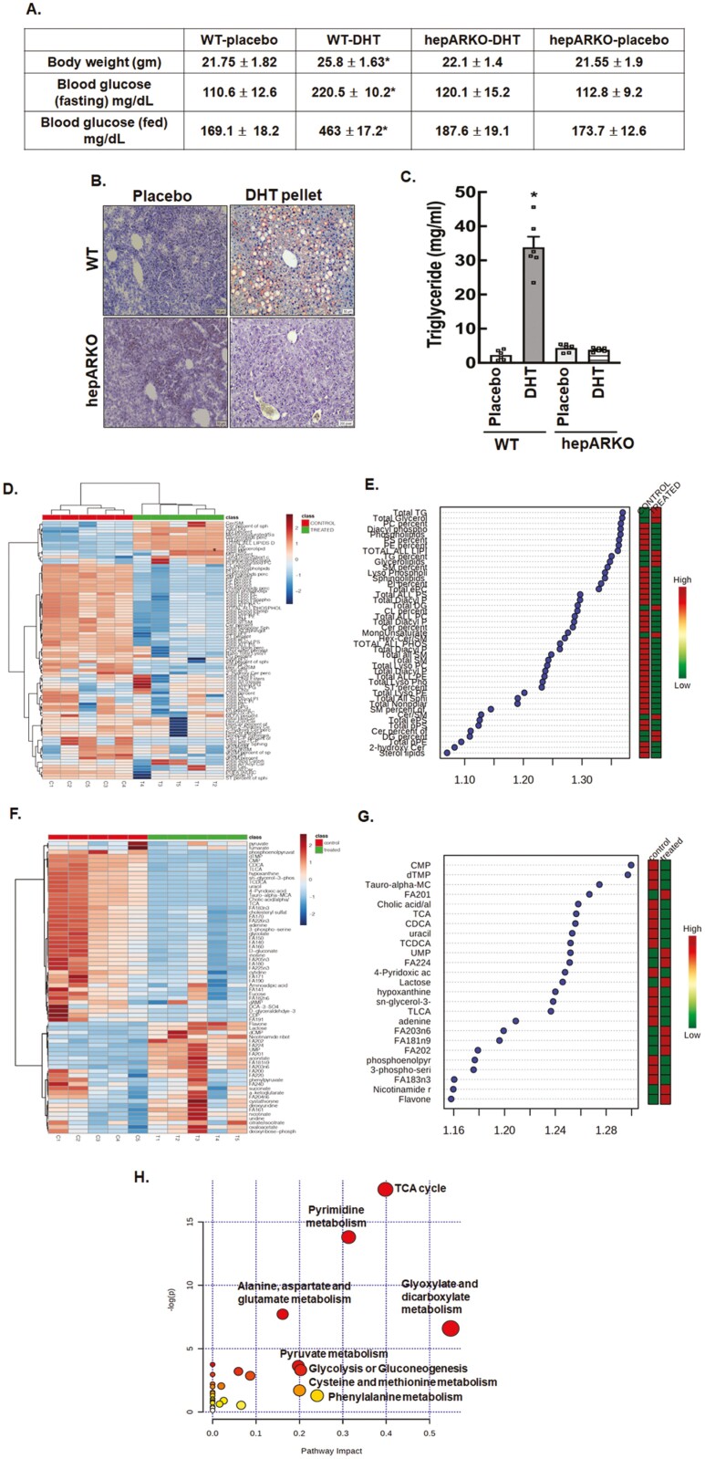
Figure 1.
Chronic high levels of androgens significantly cause hepatic steatosis. (A) Metabolic features, (B) Oil Red O staining of liver sections, and (C) liver triglyceride levels isolated from wild-type (WT) and hepatocyte-specific androgen receptor knockout (hepARKO) mice after 90 days of placebo or dihydrotestosterone (DHT) pellet treatment. Data are the mean ± SE of the mean (n = 6 mice/treatment group). *P ≤ 0.01 vs placebo using 1-way analysis of variance followed by Dunnett’s multiple comparison test). (D) Hierarchical clustering shown as heatmap of lipid classes in livers isolated from placebo (control) or DHT (treated) pellet mice (n = 5 mice/treatment). (E) Important lipid classes identified by partial least squares-discriminant analysis (PLS-DA) and represented as the average of the variable importance in projection (VIP) scores. The boxes on the right indicate the relative concentrations of the corresponding lipid class in each group (placebo-control vs DHT-treated) (F) Hierarchical clustering shown as heatmap of metabolites isolated from livers of placebo (control) or DHT (treated) pellet mice (n = 5 mice/treatment). (G) Important metabolites identified by PLS-DA and represented as the average of the VIP scores. The boxes on the right indicate the relative concentrations of the corresponding metabolites in each group (placebo control vs DHT-treated). (H) Graphical presentation of metabolome pathways in the liver impacted by DHT pellet treatment.

