. 2020 Oct 27;3(1):10–23. doi: 10.1039/d0na00454e

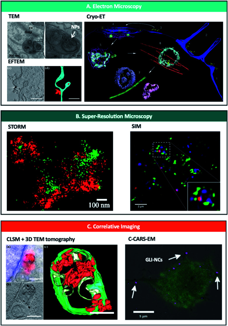
Fig. 2. Nanoscopy techniques used to study and/or quantify endo-lysosomal trafficking of nanoparticles. (A) Electron microscopy techniques including TEM (upper left),37 EFTEM (lower left)74 and Cryo-ET (right)69 can be used to track and quantify nanoparticles in intracellular vesicles. Reprinted (adapted) with permission from ref. 37 Copyright © 2013 Nature America, Inc., from ref. 74 © 2019 American Chemical Society and from ref. 69 Copyright © Azubel et al. eLife. (B) Super-resolution microscopy has been used to image nanoparticles bursting out of endosomes with STORM (left)96 and SIM (right).117 Reprinted (adapted) with permission from ref. 96 and 117 Copyright 2018 American Chemical Society. (C) Correlative imaging combines different microscopic techniques such as CLSM and 3D TEM tomography (left)74 or C-CARS and EM (right)70 and offers spatiotemporal localization of labelled NPs and biomolecules with high specificity and sensitivity at a highly subcellular level; quantification possible with CLSM + 3D TEM tomography. Reprinted (adapted) with permission from ref. 74 Copyright © 2019 American Chemical Society and from ref. 70 Copyright © 2018 The Authors. Biotechnology Journal Published by Wiley-VCH Verlag GmbH & Co. KGaA.

Fig. 2