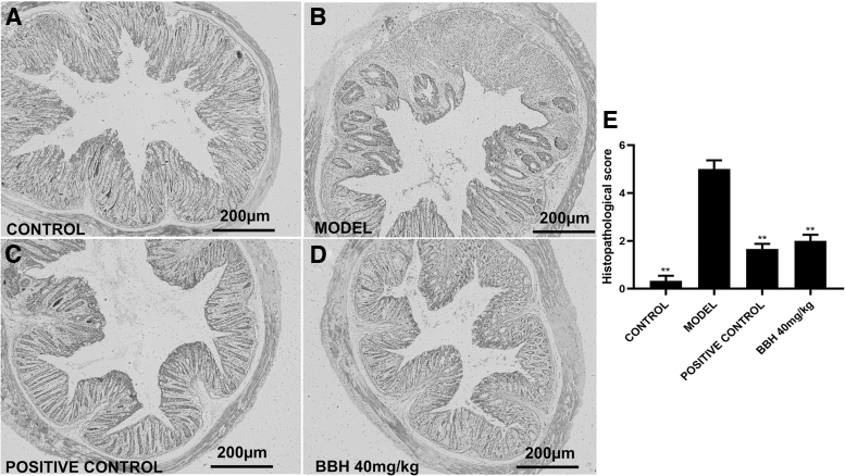
FIG. 4.
Histological features at 100 × magnification. (A) Colonic mucosa in control group with regular arranged crypts and goblet cells. (B) The histologic features in the colitis group. (C) The histologic characteristics in the SASP group. (D) The histologic characteristics in the BBH group with a dose of 40 mg/kg. (E) Histological score obtained by blind histopathological analysis. Error lines indicate means ± SEM; n = 6, **P < .01 compared with model.

