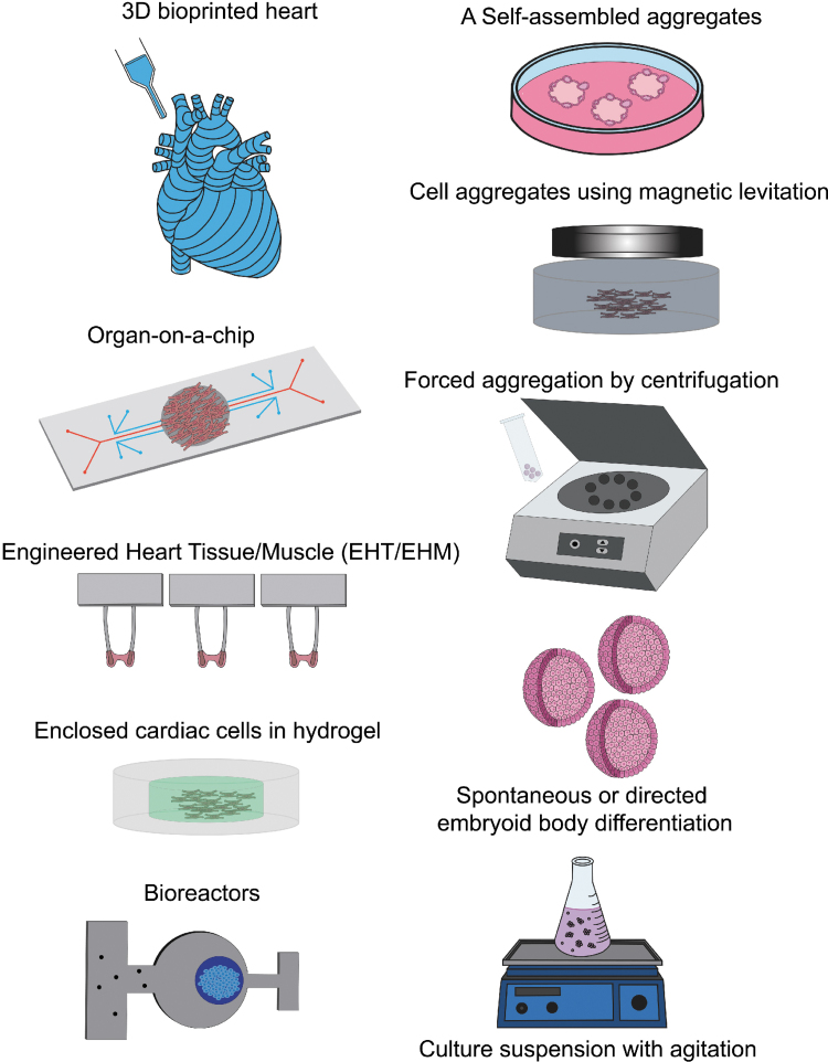
FIG. 2.
Three-dimensional induced pluripotent stem cell-derived cardiac constructs. A variety of 3D cardiac constructs have been developed to study cardiac physiology and to mimic the microenvironment of the heart. 3D bioprinted cardiac constructs promote higher spatial and anatomical accuracy with a mixture of various matrices and cardiac cells. Organ-on-a-chip and other microfluidic chips mimic complex human cardiac tissue at a miniaturized scale. EHTs/EHMs are optimized to measure the contractile force of cardiomyocytes mixed in a matrix and suspended between two posts. Hydrogels exhibit tissue-like properties to model the physiologically relevant cardiac microenvironment. Bioreactors mimic the fluid dynamics and nutrient need to assess cardiac constructs. Cardiac constructs can be generated using aggregation methods such as monolayer culture on a low adhesion surface matrix to spontaneously self-aggregate, magnetic levitation, forced aggregation using centrifugation, and spontaneous or directed cardiac differentiation in embryoid body form which can be produced at a larger scale using culture suspension with agitation (magnetic- or shaker based). Color images are available online.

