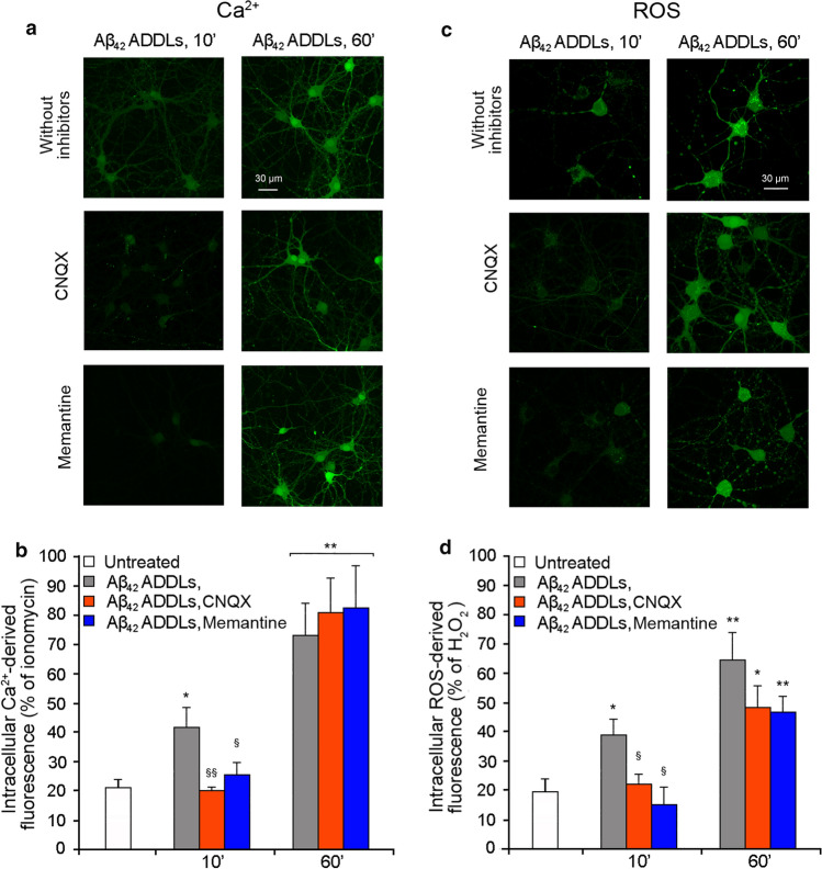
Fig. 7.
Aβ42 ADDLs oligomers increase intracellular Ca2+ levels and ROS production in primary rat cortical neurons. a Representative confocal scanning microscopy images of intracellular free Ca2+ levels in primary rat cortical neurons treated with no inhibitors (first row), 5 µM CNQX (second row) and 10 µM memantine (third row), and analysed after 10 and 60 min of treatment with 1 µM (monomer equivalents) Aβ42 ADDLs oligomers. b Semi-quantitative analysis of intracellular free Ca2+-derived fluorescence. c Representative confocal scanning microscopy images of intracellular ROS levels in primary rat cortical neurons treated with no inhibitors (first row), 5 µM CNQX (second row) and 10 µM memantine (third row), and analysed after 10 and 60 min of treatment with 1 µM (monomer equivalents) Aβ42 ADDLs oligomers. d Semi-quantitative analysis of intracellular ROS-derived fluorescence. Three different experiments were carried out, with 10–22 cells each, for each condition. Data are represented as mean ± SEM (n = 3). The single (*) and double (**) asterisks refer to p values < 0.05 and < 0.01, respectively, relative to untreated cells. The single (§) and double (§§) symbols refer to p values < 0.05 and < 0.01, respectively, relative to Aβ42 ADDLs oligomers without inhibitors at corresponding time points

