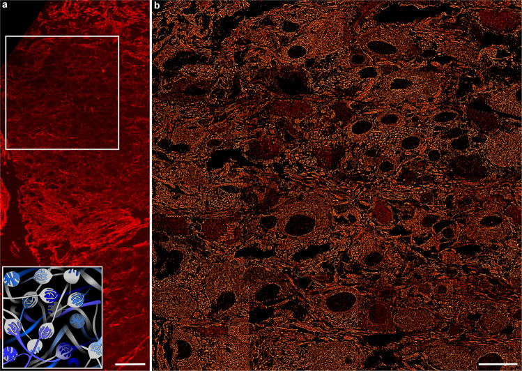
Fig. 5. Accelerated large-sample imaging with DeepSTORM DNA-PAINT.
a Confocal microscopy image of an MNTB tissue section and a graphical representation of calyces organised within the MNTB region (inset; blue indicates principal cells and in grey are the postsynaptic Calyx of Held). b Large-area super-resolution image recorded for the tissue area defined by the bounding box in (a). The α-tubulin super-resolution image was obtained by imaging 55 µm × 55 µm patches recorded with 10 nM imager strand P1 in a 4 × 4 grid-like fashion with 400 frames per patch, obtaining high-density DNA-PAINT frames in 1 min per image and a total imaging time of 16 min. N = 1 tissue sample; scale bar 50 µm (a), 20 µm (b).

