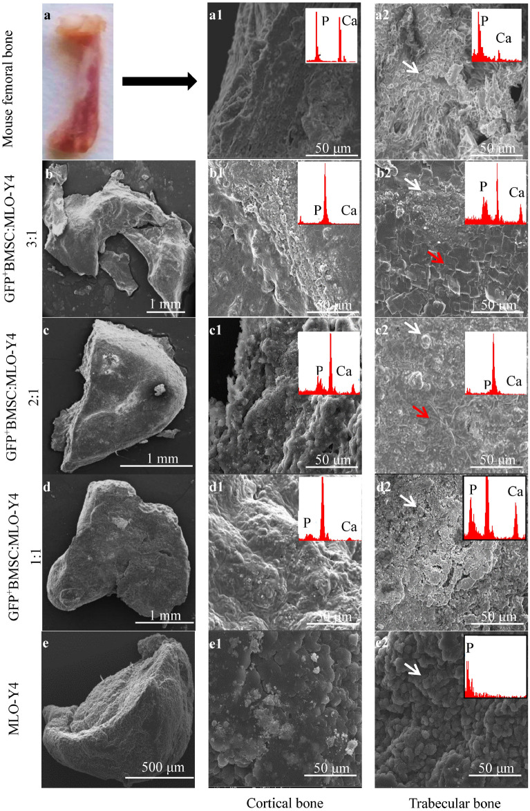
Figure 2.
SEM morphology of the cross-sections of bone-like tissues and mouse femoral bone. Continuous dense layers of deposited calcium phosphate and ECMs were observed in the mouse femoral cortical bone (a1) and with GFP+BMSC:MLO-Y4 3:1 (b1), 2:1 (c1), and 1:1 (d1), but not with the MLO-Y4 monoculture (e1). Spherical calcium phosphate was observed in mouse femoral trabecular l bone (a2) and all of the spheroid cultures (b2–e2, white arrowheads). Flake-like calcium phosphate was present in the 3:1 (b2) and 2:1 (c2) co-cultures (red arrowheads). SEM, scanning electron microscopy.

