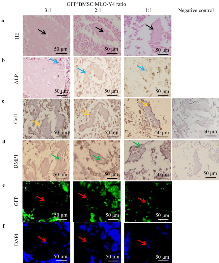
Figure 3.
The histomorphological and immunohistochemical features of the bone-like tissues. (a) HE staining showed osteoblast-like cells surrounded by a cohesive osteoid-like matrix and osteocyte-like cells embedded into lacunae-like structures (black arrowheads) in the 3:1, 2:1, and 1:1 co-cultures. (b,c) Osteoblast-like cells on the osteoid-like matrix surface were positive for ALP (blue arrowheads) and Col1 (orange arrowheads). (d) Osteocyte-like cells in the osteoid-like matrix were positive for DMP1 (green arrowheads). (e,f) Fluorescence microscopy showed that osteocyte-like cells embedded in the osteoid-like matrix and osteoblast-like cells on the surface were GFP positive (red arrowheads). DAPI, 4′,6-diamidino-2-phenylindole.

