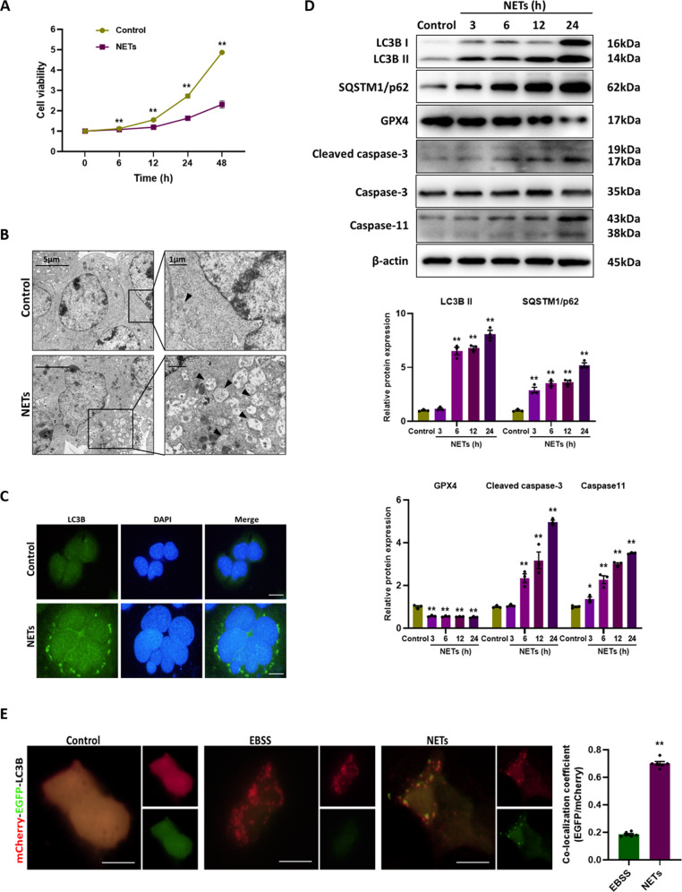
Fig. 4. NETs impair the cell viability via inducing autophagosome formation but blocking autophagic flux.
A The cell viability of alveolar epithelial cells with different treatments. B Representative electron microscopic images of autophagic vesicles (black arrow) in alveolar epithelial cells with different treatments. C Representative images of immunofluorescence staining of LC3B in alveolar epithelial cells with different treatments. Scale bar: 20 µm. D Western blot images of autophagy signaling and cell death markers expression in alveolar epithelial cells. E Representative fluorescent images of alveolar epithelial cells transfected with mCherry-EGFP-LC3B and treated with EBSS or NETs. Scale bar: 10 µm. Each bar shows means ± SEM. The comparison between two groups was conducted by unpaired t-test. *p < 0.05, **p < 0.01 versus the control group.

