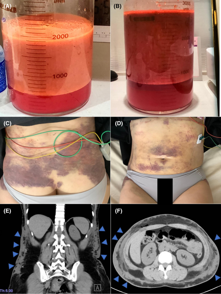
Fig. 1.

(A) The collected fluids at the first surgery. (B) The collected bloody fluids at the secondary surgery, which was bloodier than the previous one. (C,D) Diffuse purpuras were identified around the abdomen. (E,F) Computed tomography showing diffused hematomas all over the abdomen (arrowheads).
