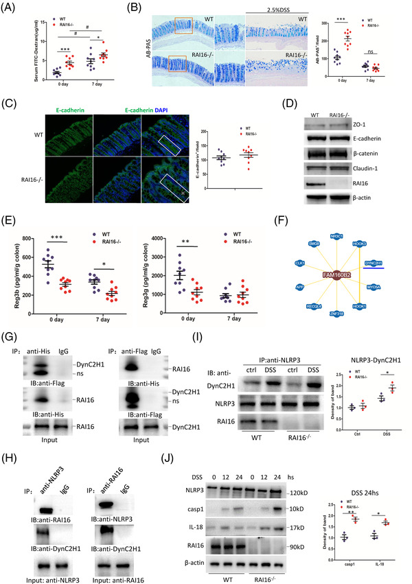
FIGURE 2.

RAI16 interacts with DynC2H1 regulating DSS‐induced NLRP3 activation. (A) Intestinal permeability of WT and RAI16–/– mice (n = 9) was determined with FITC‐dextran in vivo. (B) Alcianblue‐Periodic acid Schiff (AB‐PAS; goblet cells) staining of the colons derived from WT and RAI16–/– mice (n = 9) treated with or without 2.5% DSS for 6 days and quantitation results are shown in the right. (C) Representative E‐cadherin staining in colon sections derived from WT and RAI16–/– mice (n = 9) and quantitation results are shown in the right. (D) Immunoblotting analysis of the expression of selected adherent junction (AJ) and tight junction (TJ) proteins in colonic epithelial scrapings obtained from WT and RAI16–/– mice. (E) Reg3β and Reg3γ levels from WT and RAI16–/– colon tissue explants were measured by ELISA. (F) The predict interaction of RAI16 (Fam160B2) and DynC2H1. (G) HEK293T cells were co‐transfected with RAI16‐His and DynC2H1‐Flag expression vectors. The cell lysates were immunoprecipitated using anti‐Flag or anti‐His and immunoblotted with anti‐His or anti‐Flag. (H) Endogenous co‐immunoprecipitation assay was performed in IECs with anti‐NLRP3 or anti‐ RAI16, and detected with indicated antibodies. (I) IECs treated with DSS (1.0%) or not for 24 h were immunoprecipitated with anti‐DynC2H1, anti‐RAI16 or IgG, and detected with indicated antibodies. (J) IECs from WT and RAI16–/– mice were treated with DSS and immunoblotted with indicated antibodies.
