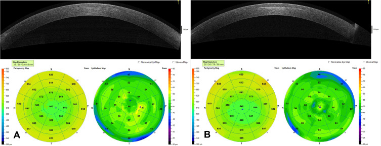
Figure 10.
This image contains a corneal OCT and corneal topography 9 months after implantation of Raindrop inlay showing normal appearance of inlay in corneal stroma (A). Corneal OCT and topography 4 months after explantation of the Raindrop inlay demonstrating significant residual haze at the former site of the inlay in the OCT scan and changes in topography (B). (Courtesy of Hoopes Vision).

