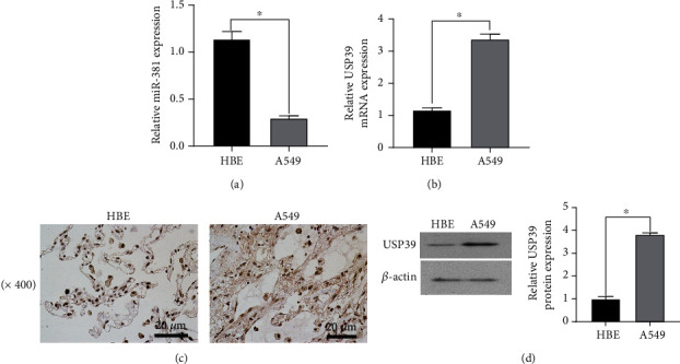
Figure 1.

Expression of miR-381 and USP39 in tissues and cell lines of non-small-cell lung cancer. (a) The left figure shows the detection of miR-381 in different lung cancer tissues and adjacent normal tissues by qRT-PCR. The right image shows the expression level of miR-381 in different lung cancer tissues. ∗p < 0.05 versus normal tissues, versus HBE. (b) Expression of USP39 in lung cancer and adjacent normal tissues. ∗p < 0.05 versus normal tissues. (c) Immunohistochemical detection of USP39 expression in lung cancer cells and normal tissues. (d) WB detection of USP39 expression in lung cancer cells and normal tissues. ∗p < 0.05 versus normal tissues.
