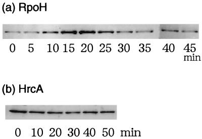
FIG. 8.
The cellular levels of RpoH and HrcA during the heat shock response. Cells of wild-type A. tumefaciens (GV3101) were grown to mid-log phase in complete medium at 25°C and shifted to 37°C at time zero. Samples were taken at the indicated times, mixed with an equal volume of 2× sodium dodecyl sulfate (SDS) loading buffer, and boiled for 5 min. Equal amounts of protein adjusted by the optical density (in Klett units) of the culture were loaded on the SDS-polyacrylamide gels (12.5% gels), blotted onto a nitrocellulose membrane (Hybond ECL; Amersham Life Science), and detected with rabbit antiserum against RpoH (a) or HrcA (b) by chemiluminescence techniques.

