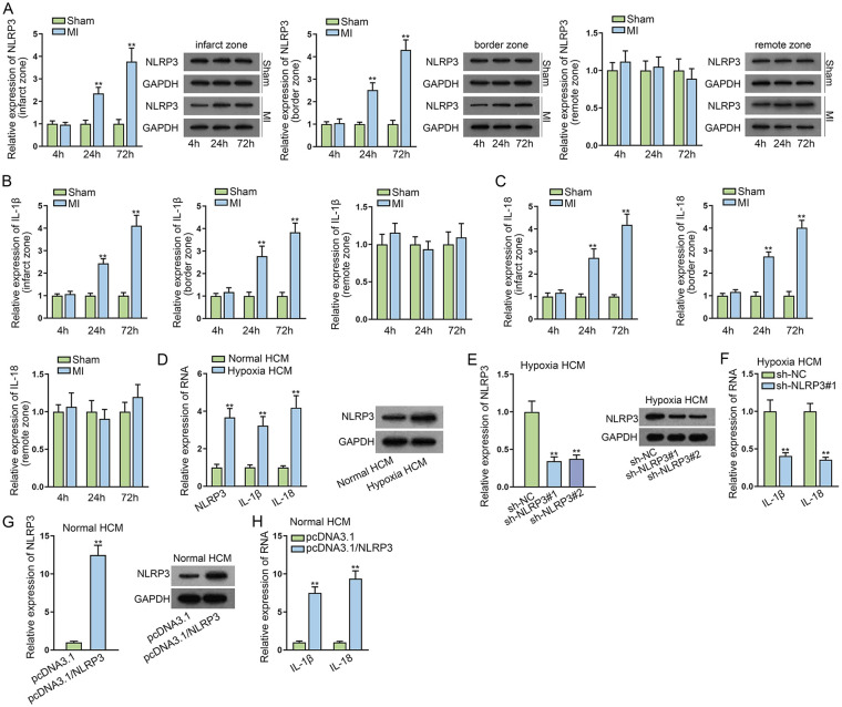
Figure 1.
NLRP3 is overexpressed in mice MI models and hypoxia-induced myocardial cells. (A) RT-qPCR and WB detected the NLRP3 level in infarct zone, border zone, and remote zone after MI for 4, 24, and 72 h, respectively. (B, C) RT-qPCR and WB measured IL-1β and IL-18 expression in infarct zone, border zone, and remote zone after MI for 4, 24, and 72 h, respectively. (D) The expression of NLRP3, IL-1β, and IL-18 was assessed in hypoxia HCMs compared with the normal group through RT-qPCR and WB. (E) The expression of NLRP3 was detected in hypoxia HCMs transfected with shRNAs targeting NLRP3 via RT-qPCR and WB. (F) RT-qPCR examined the expression of IL-1β and IL-18 after NLRP3 silencing in hypoxia HCMs. (G) RT-qPCR and WB evaluated NLRP3 level in normal HCMs transfected with pcDNA3.1/NLRP3. (H) The expression of IL-1β and IL-18 was examined in normal HCMs transfected with pcDNA3.1/NLRP3 using RT-qPCR. HCM: human cardiomyocytes; MI: myocardial infarction; NLRP3: NLR family pyrin domain containing 3; NLRP3: NLR family pyrin domain containing 3; RT-qPCR: reverse transcription quantitative polymerase chain rection; WB: western blot. **P < 0.01.

