
Figure 1.
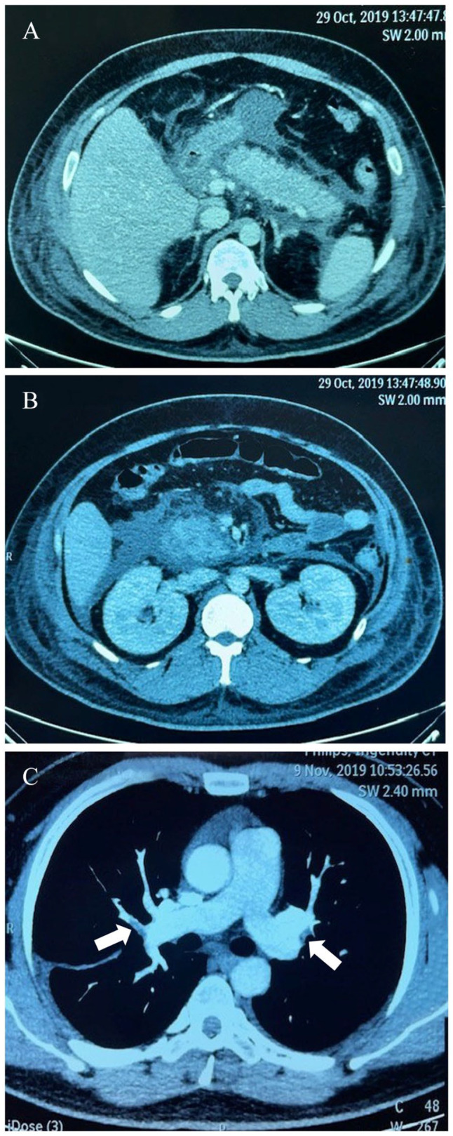
Computed tomography scan of the abdomen revealed: (A) diffuse swollen pancreas with surrounding retroperitoneal fat stranding, (B) liquefactive necrosis of pancreatic parenchyma, and (C) CT angiography revealed intraluminal filling defects (arrowhead) in subsegmental pulmonary arteries, compatible with acute pulmonary embolism.