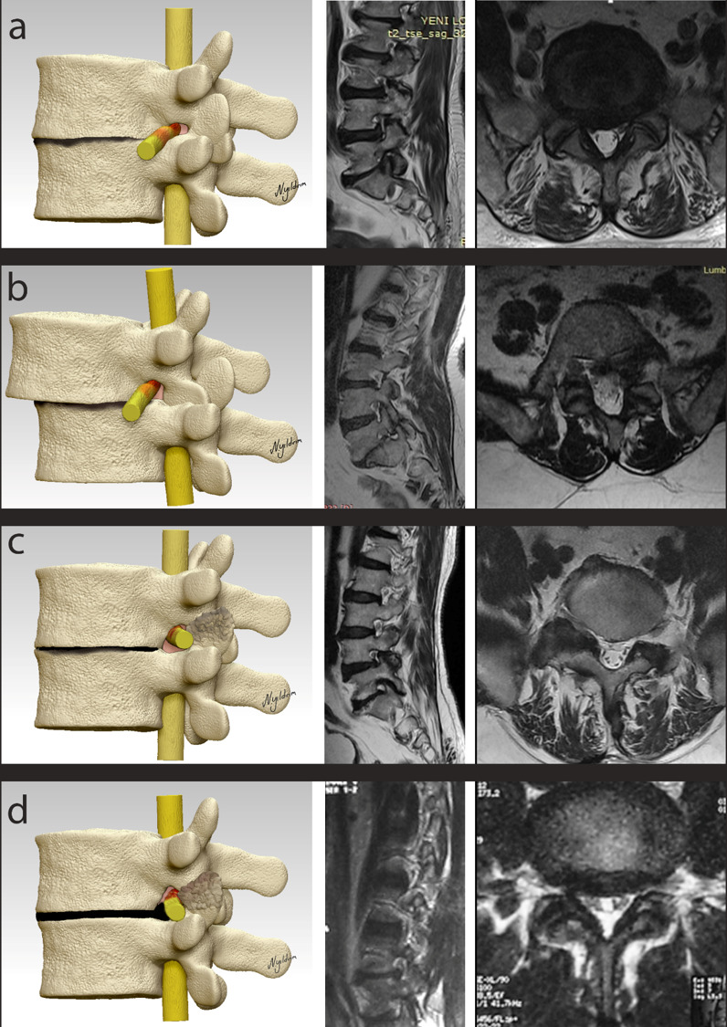
Figure 2.

(a) Type I stable foraminal stenosis. The disc has completely degenerated; the root is squeezed between the upper and lower pedicles. (b) Type II stable foraminal stenosis. The disc has completely degenerated at the base of degenerative spondylolisthesis. The root is squeezed between the upper pedicles and lower vertebral corpus. (c) Type III stable foraminal stenosis. The disc has completely degenerated; the root is squeezed between the calcified facet joint and the corner of the upper pedicles and posterior wall of upper vertebrae. (d) Type IV stable foraminal stenosis. The disc is calcified in some areas and fused in some areas to the upper and lower vertebral corpus, with bulging of the posterior annulus. The root is squeezed between the calcified bulging of the posterior annulus, calcified facet joint, and upper pedicles. For all images, the red areas on the nerve root indicate dense areas where the nerve is squeezed.
