Figure 2.
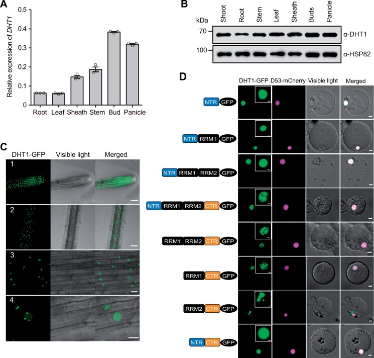
DHT1 gene is widely expressed in plant tissues and the DHT1 protein is localized in nuclear speckles. A, RT-qPCR analysis showing DHT1 transcript levels in different rice tissues. Data are means ± sem (n = 3). B, Immunoblot analysis showing DHT1 protein levels in different tissues. C, Nuclear localization of DHT1-GFP(green fluorescent protein) fusion protein in transgenic rice root cells. Number 1 shows the cell division region in roots; number 2 shows the cell maturation region in roots; numbers 3 and 4 are magnified images of the cell maturation region in roots. Scale bars: 50 μm (1, 2), 20 μm (3), and 10 μm (4). D, The subcellular localization of different domains of DHT1 protein in rice protoplasts. Scale bars, 5 μm. The top right inset images show the magnified green fluorescent portions of DHT1-GFP. Scale bars, 2 μm.

