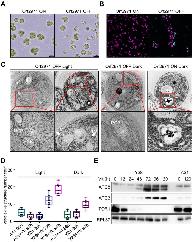
Figure 3.
Autophagy-like phenotype after the depletion of Orf2971. A, Light microscopy of Y28 cells in the presence (Orf2971 OFF) or absence of vitamins (Orf2971 ON). B, Y28 cells treated or untreated with vitamins were stained with autophagy indicator MDC (Dansylcadaverine) to visualize autophagosomes. Cells were imaged by confocal microscopy. The merged channels of chlorophyll fluorescence and MDC stain fluorescence are shown. Cells were grown in the light for 96 h. C, Analysis by transmission electron microscopy of epoxy-embedded thin sections of Y28 (Orf2971 OFF) and A31 (Orf2971 ON) cells following treatment by vitamins for 72 and 96 h. The images in the second row represent the enlarged area in the rectangle of the first row. V, vacuoles; RCB, double membrane Rubisco-containing bodies; TM indicates thylakoid membranes engulfed in the autophagy-like vesicles and Py indicates the pyrenoid. D, Number of vacuoles and vesicle-like structures in A31 and Y28 in the presence or absence of vitamins. Data are represented as mean ± sd. E, Autophagy-related proteins were examined at different time points after addition of vitamins by immunoblotting using antibodies against the indicated proteins.

